Japanese Classification of Esophageal Cancer 11th Edition. And in one patient with to and fro the flow of LGV was not demonstrated.

Organ Related Staging And Grading Abdominal Key
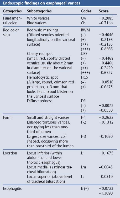
General rules for recording endoscopic findings of esophageal varices were initially proposed in 1980 and revised in 1991.

. D Large white oesophageal varices that look like mucosal folds. 72 Classification of esophageal varices. 2007 the 11th edition of the Japanese Classification of Esophageal Cancer has now been published.
In addition following the adoption of cri-teria for the diagnosis of lesions located at the. Part II and III. The present paper evaluates the usefulness of ECE for diagnosing EV in Japanese patients with cirrhosis.
General rules for recording endoscopic findings of esophageal varices were initially proposed in 1980 and revised in 1991. Small low-risk esophageal varices or F1 almost flattened out with insufflation. A Small oesophageal varices grade 1 form F1.
We examined 29 patients with cirrhosis 20 males and 9. These rules have widely been used in Japan and other countries. Although esophageal capsule endoscopy ECE is reportedly useful in the diagnosis of esophageal varices EV few reports have described the benefits of this technique in Asian countries.
Recently portal hypertensive gastropathy has been recognized as a distinct histological and functional entity. However since the development of endoscopic sclerotherapy and other modalities of endoscopic treatment these 1980 rules were found to be insufficient for recording mucosal changes after treatment. Gastroesophageal varices GOV are varices that extend from the esophagus to the stomach and can be classified into 2 groups.
Several studies have shown that some endoscopic features of esophageal varices such as large size and the presence of red signs on their surface are associated with the risk of variceal hemorrhage. Although esophageal capsule endoscopy ECE is reportedly useful in the diagnosis of esophageal varices EV few reports have described the benefits of this technique in Asian countries. 1 GOV 1 varices are continuous with esophageal varices and extend along the lesser curve for about 2 to 5 cm below the gastro-esophageal junction and 2 GOV2 varices extend from the esophagus below the gastro-esophageal junction toward the.
Large esophageal varices larger than one third of the esophageal lumen with some high-risk stigmata red marks or wheals. F Large oesophageal varices with red colour signs cherry. C Large oesophageal varices grade 3 form F3.
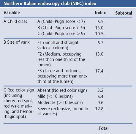
Epub 2016 Nov 10. A prognostic index that uses these endoscopic signs in association with an index of liver function such as the Childs classification has been shown to be useful in. They are most often a consequence of portal hypertension commonly due to cirrhosis.
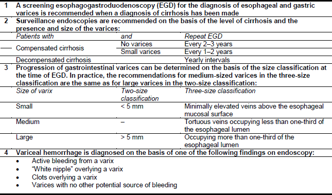
Part II and III Esophagus. The North-Italian Endoscopic Club NIEC used a similar definition ie small medium and large corresponding to a variceal diameter of 066 of the radius of the esophagus respectively Fig. The sensitivity specificity positive predictive value and negative predictive value of M2BPGi with a cutoff value of 5 COI was 926 701 556 and 959 respectively.
Endoscopic ultrasonography can clearly depict vascular structures around the. I20 Both groups take into account the size of the largest varix seen in the esophagus. The general rules as revised in.
Japanese Classification of Esophageal Cancer 11th Edition. Of these flow direction of LGV changed from hepatofugal to hepatopetal in two from hepatofugal to to and fro in two. After ESL esophageal varices were eradicated in five patients endoscopically.
National Center for Biotechnology Information. B Small and beady oesophageal varices grade 2 form F2. Endoscopic ultrasonography can clearly depict vascular structures around the.
These rules have widely been used in Japan and other countries. The present paper evaluates the usefulness of ECE for diagnosing EV in Japanese patients with cirrhosis. The revised Japanese classification by the Italian Liver Cirrhosis Project ILCP group describes variceal size as the percentage of radius of the esophageal lumen occupied by the largest varix8 A small or grade 1 varix is said to occupy less than 25 of the lumen a medium or grade 2 varix occupies 25 to 50 of the lumen and a large or grade 3 varix occupies.
Medium-sized low-risk esophageal varices F2 that did not flatten with insufflation. Recently portal hypertensive gastropathy has been recognized as a distinct histological and functional entity. E Grade 3 oesophageal varices with red colour signs whip-like red wale marks.
The general rules made in 1980 for recording endoscopic findings of esophageal varices have widely been used in Japan and in other countries. Esophageal varices are extremely dilated sub-mucosal veins in the lower third of the esophagus. People with esophageal varices have a strong tendency to develop severe bleeding which left untreated can be fatal.
During this period supplements to the 10th edition involving the revision of disease typing and terminology were pub-lished in 2008. Esophageal varices are typically diagnosed through an. We examined 29 patients with cirrhosis 20 males and 9.

Endoscopic Classification Of Esophageal Varices According To The Download Table

Wgo Esophageal Varices Guideline Summary

Endoscopic Management Of Esophagogastric Varices In Japan Semantic Scholar

Table 1 From Esophageal Capsule Endoscopy For Screening Esophageal Varices Among Japanese Patients With Liver Cirrhosis Semantic Scholar

Endoscopic Classification Of Esophageal Varices According To The Download Table

Clinical Outcomes Of Gastric Variceal Obliteration Using N Butyl 2 Cyanoacrylate In Patients With Acute Gastric Variceal Hemorrhage

Prediction Of The First Variceal Hemorrhage In Patients With Cirrhosis Of The Liver And Esophageal Varices Nejm

Prediction Of The First Variceal Hemorrhage In Patients With Cirrhosis Of The Liver And Esophageal Varices Nejm

Table 1 From Correlation Between Serum Ascites Albumin Concentration Gradient And Endoscopic Parameters Of Portal Hypertension Semantic Scholar

Table 1 From Correlation Between Serum Ascites Albumin Concentration Gradient And Endoscopic Parameters Of Portal Hypertension Semantic Scholar

Wgo Esophageal Varices Guideline Summary

Endoscopic Classification Of Esophageal Varices According To The Download Table

Japanese Classification Of Esophageal Varices Benytr

Pdf Esophageal Capsule Endoscopy For Screening Esophageal Varices Among Japanese Patients With Liver Cirrhosis Semantic Scholar

Endoscopic Classification Of Esophageal Varices According To The Download Table

Endoscopic Diagnosis Grading And Predictors Of Bleeding In Esophageal And Gastric Varices Techniques In Gastrointestinal Endoscopy


